


by
, , , , , , , , , , , and

Cells 2023, 12(16), 2027; https://doi.org/10.3390/cells12162027 - 08 Aug 2023
Abstract
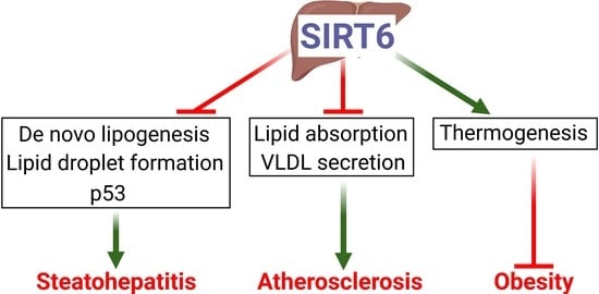
Although very common, the precise mechanisms that explain the symptomatology of neuroendocrine syncope (NES) remain poorly understood. This disease, which can be very incapacitating, manifests itself as a drop in blood pressure secondary to vasodilation and/or extreme slowing of heart rate. As studies
[...] Read more.
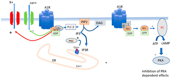
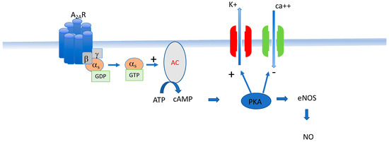
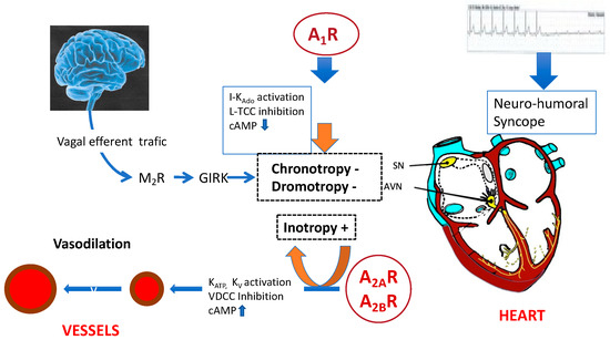
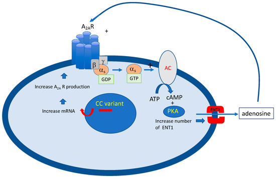
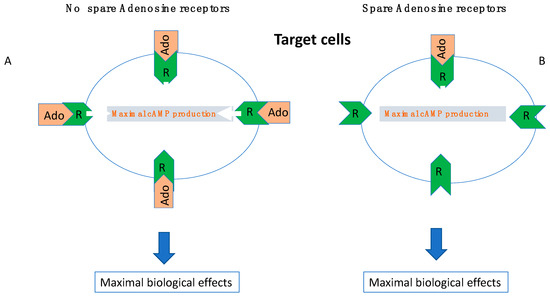
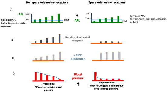
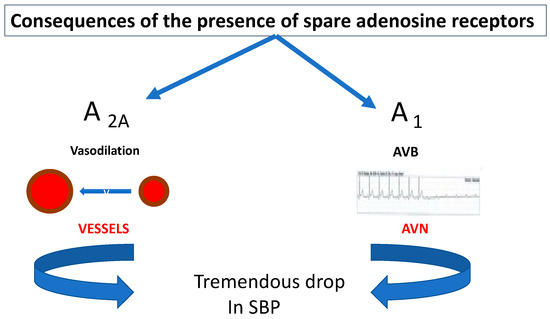
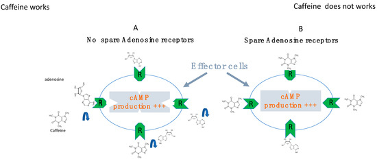
Although very common, the precise mechanisms that explain the symptomatology of neuroendocrine syncope (NES) remain poorly understood. This disease, which can be very incapacitating, manifests itself as a drop in blood pressure secondary to vasodilation and/or extreme slowing of heart rate. As studies continue, the involvement of the adenosinergic system is becoming increasingly evident. Adenosine, which is an ATP derivative, may be involved in a large number of cases. Adenosine acts on G protein-coupled receptors with seven transmembrane domains. A1 and A2A adenosine receptor dysfunction seem to be particularly implicated since the activation leads to severe bradycardia or vasodilation, respectively, two cardinal symptoms of NES. This mini-review aims to shed light on the links between dysfunction of the adenosinergic system and NHS. In particular, signal transduction pathways through the modulation of cAMP production and ion channels in relation to effects on the cardiovascular system are addressed. A better understanding of these mechanisms could guide the pharmacological development of new therapeutic approaches.
Full article
(This article belongs to the Special Issue Adenosine and Adenosine Receptors in Human Disease)
►
Show Figures
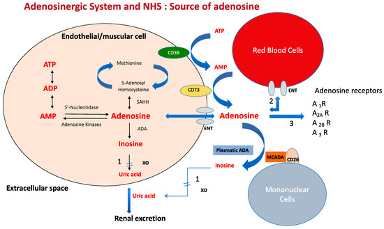
Figure 1


.jpg)


